
Chapter 8
The Cricket Gryllus bimaculatus:
Techniques for Quantitative and Functional
Genetic Analyses of Cricket Biology
Arpita Kulkarni and Cassandra G. Extavour
Abstract All extant species are an outcome of nature’s “experiments” during
evolution, and hence multiple species need to be studied and compared to gain a
thorough understanding of evolutionary processes. The field of evolutionary devel-
opmental biology (evo-devo) aspires to expand the number of species studi ed,
because most functional genetic studies in animals have been limited to a small
number of “traditional” model organisms, many of which belong to the same phylum
(Chordata). The phylum Arthropoda, and particularly its component class Insecta,
possesses many important characteristics that are considered favorable and attractive
for evo-devo research, including an astonishing diversity of extant species and a
wide disparity in body plans. The development of the most thoroughly inves tigated
insect genetic model system to date, the fruit fly Drosophila melanogaster
(a holometabolous insect), appears highly derived with respect to other insects and
indeed with respect to most arthropods. In comparison, crickets (a basally branching
hemimetabolous insect line age compared to the Holometabola) are thought to
embody many developmental features that make them more representative of
insects. Here we focus on crickets as emerging model s to study problems in a
wide range of biological areas and summarize the currently available molecular,
genomic, forward and reverse genetic, imaging and computational tool kit that has
been establis hed or adapted for cricket research. With an emphasis on the cricket
species Gryllus bimaculatus, we highlight recent efforts made by the scientific
community in establishing this species as a laboratory model for cellular biology
and developmental genetics. This broad toolkit has the potential to acceler ate many
traditional areas of cricket research, including studies of adaptation, evolution,
A. Kulkarni
Department of Organismic and Evolutionary Biology, Harvard University, Cambridge, MA,
USA
C. G. Extavour (
*)
Department of Organismic and Evolutionary Biology, Harvard University, Cambridge, MA,
USA
Department of Molecular and Cellular Biology, Harvard University, Cambridge, MA, USA
e-mail: [email protected]
© Springer Nature Switzerland AG 2019
W. Tworzydlo, S. M. Bilinski (eds.), Evo-Devo: Non-model Species in Cell and
Developmental Biology, Results and Problems in Cell Differentiation 68,
https://doi.org/10.1007/978-3-030-23459-1_8
183
neuroethology, physiology, endocrinology, regeneration, and reproductive behavior.
It may also help to establish newer areas, for example, the use of crickets as animal
infection model systems and human food sources.
8.1 Introduction
All cellular life forms share a last common ancestor. Every extant species is a current
outcome of an evolutionary process that has taken place over hundreds of millions of
years. Thus, each species that exists today can be used as a data point toward
increasing our understanding of the living world. Comparative study of the devel-
opmental biology of multicellular organisms motivates the field of evolutionary
developmental biology, or “evo-devo.” However, to date only a relatively small
number of species has been used to study the functions of genes that regulate
animal developmental processes, which limits our understanding of how develop-
mental genetic changes underpin evolution. If the idealized goal of evo-devo
research is to do a speci es comparison that includes representatives of all major
evolutionary transitions, then this calls for the establishment of many more animals
as laboratory model organisms than is currently the case. As a step in this direction,
developmental biologists are increasingly choosing new animal models that are
suitable to address thus far neglected areas of research, while sim ultaneously
selecting these candidates for their ability to fulfill other criteria relevant for
evo-devo work. Some criteria that could maximize the scientific gains achieved by
establishing new models include choosing organisms that belong to clades that are
representative of a wide range of ecolog ical niches, and those belonging to phyla that
are species-rich, display high diversity in form and function, have interdisciplinary
scientific appeal, are economical to maintain in the laboratory, and could inform
issues that directly affect humans (e.g., disease or agriculture). Strategically choos-
ing and studying examp les satisfying some or all of these criteria may therefore be
impactful, evolutionarily informative, and a good use of limited resour ces.
The phylum Arthropoda contains multiple species that satisfy many of the above
criteria and has thus played a prominent role in modern evo-devo research. Impor-
tantly, good fossil records exist for this phylum, providing researchers with snap-
shots into the evolutionary past and aiding in comparative work. For example, the
EDNA fossil insect database lists over 23,000 species (Giribet and Edgecombe
2013; Mitchell 2013), with the earliest records of arthropod fossils dating back to
nearly 555 million years ago (Mya) during the Cambrian era (Harvey et al. 2012;
Zhang et al. 2010; Vaccari et al. 2004). Undoubtedly, access to such fossil records is
essential to understanding the key phenotypic innovations that have made
Arthropoda species-rich and evolutionarily successful (Mayhew 2007).
Extensive studies on insects, which account for the majority of all species
described on earth (Wheeler 1990; Grimaldi and Engel 2005) and for ~85% of all
arthropod diversity (Giribet and Edgecombe 2013), have pioneered and shaped the
evo-devo field. This work has informed us of the genetic and evolutionary basis of
184 A. Kulkarni and C. G. Extavour
pivotal developmental mechanisms. For example, the description of the first home-
otic mutant (Bridges and Morgan 1923), and the realization of the significance of
conserved developmental genes in body patterning and in the evolution of different
body plans across animals, come from studies in the insect Drosophila melanogaster
(Lewis 1978; Nüsslein-Volhard and Wieschaus 1980; Duboule and Dolle 1989;
Graham et al. 1989 ; Panganiban et al. 1997; Heffer et al. 2013). Other insect-based
research that has broadened our understanding of evolutionary processes includes
work on key evolutionary innovations such as the insect body plan (Grimaldi and
Engel 2005), metamorphosis (Truman and Riddiford 1999), development of wings
(Nicholson et al. 2014; Alexander 2018; Bruce and Patel 2018 ; Linz and Tomoyasu
2018), morphological novelties (Kijimoto et al. 2013), and insect eusociality (Toth
and Rehan 2017). The widespread scope of such research is a result of the practical
advantages that come with working on insects: insect phylogeny is well established
(Giribet and Edgecombe 2013), many species are easy to mai ntain and culture in the
laboratory, are often amenable to functional genetic analysis, in many instances
produce easily accessible large broods suitable for external manipulation, and often
have life cycles sufficiently short to allow laboratory rearing and multigenerational
analysis.
To date, popular insect models to study the genetic basis of development have
included the fruit fly Drosophila melanogaster (Diptera) (Demerec 1950), the flour
beetle Tribolium castaneum (Coleoptera) (Sokoloff 1966, 1972, 1974, 1977; Denell
2008), the honeybee Apis mellifera (Hymenoptera) (Gould and Grould 1995;
Oldroyd and Thompson 2006), the wasp Nasonia vitripennis (Hymenoptera)
(Werren and Loehlin 2009), and the silk moth Bombyx mori (Lepidoptera) (Xia
et al. 2004; Goldsmith et al. 2005; Meng et al. 2017). All of these insects, however,
share a commonality: they belong to the same insect superorder of Holometabola, or
insects that undergo complete metamorphosis during development. Complete meta-
morphosis is characterized by a pupal stage in the transition from larvae to adults,
with neither the larval nor the pupal stages resembling the final adult form (Truman
and Riddiford
1999). Such insects display a number of evolutionarily derived
developmental characters that are not generally representative of all insects, let alone
all arthropods (Mito and Noji 2008). To correct the overrepresentation of holome-
tabolous insects in modern comparative developmental literature, insects branching
basally to the Holometabola should be studied, as they, based on parsimony, appear
to display characters that are likely ancestral to insects and in some cases also to
arthropods. These insects are the Hemimetabola, insects displaying incomplete
metamorphosis and lacking pupal stages during development. The embryo in such
insects develops into a miniature adult (referred to as a nymph or a juvenile) which
then undergoes several successive molts before reaching a dulthood and sexual
maturity. Orthopterans (crickets, grasshoppers, and locusts), one of the most abun-
dant and dominant terrestrial insect groups, are in this category (Grimaldi and Engel
2005). Orthopterans display extraordinary diversity in developmental processes and
are also economical ly important herbivores, which has resulted in them becoming
popular for functional genetic research in recent years.
8 The Cricket Gryllus bimaculatus: Techniques for Quantitative .. . 185
For the rest of this chapter, we focus on crickets as promising evo-devo models,
deserving of serious regard. We discuss Gryllus bimaculatus,afield cricket species,
introduce this model system, and discuss recent advances in estab lishing this animal
for cell, developmental, and genetic research.
8.2 A Hemimetabolous Insect Model: The Cricket Gryllus
bimaculatus De Geer
G. bimaculatus is a cosm opolitan orthopteran belonging to the family Gryllidae and
is, to our knowledge, the most widespread of all Gryllus specie s for laboratory
studies (Otte and Cade 1984). Although its use for developmental genetics is
relatively recent, this species is by no means new to biological research:
G. bimaculatus has been extensively used to inform areas such as neurobiology ,
insect physiology, reproduction, and behavior since the 1960s (Huber et al. 1989;
Engel and Hoy 1999; Paydar et al. 1999; Wenzel and Hedwig 1999; Hedwig and
Poulet 2004; Nakamura et al. 2008a, b; Horch et al. 2017b). The discovery of RNA
interference (RNAi) as a mechanism for abrogating gene function (reviewed by Sen
and Blau 2006) has greatly accelerated G. bimaculatus research (Mito et al. 2011),
yielding important information about the developmental biology of this organism
and unveiling its potential as an upcoming functional genetics laboratory model.
This specie s was first described by Baron Charles de Geer in 1773 (Geer 1773)
and named Gryllus (meaning “ cricket” in Latin) bimaculatus (from the Latin “mac-
ula” for “spot ” ). Indeed, this species is commonly referred to as the “two spotted
field cricket,” for the white spot that this species displays on the dorsal surfa ce of the
forewings next to the pronotal margin (Fig. 8.1a) (Otte and Cade 1984). Some of the
morphological keys used to distinguish adult G. bimaculatus from other similar
looking field cricket speci es, both within and outside the genus Gryllus (Otte and
Cade 1984), include the white spots, a black colored adult body size of ~3 0 mm
lacking any bands, forewings nearly covering and large hindwings extending well
beyond the abdomen, and an ovipositor (in females) slightly longer than the hind
femora (Fig. 8.1a). As an additional tool in the field, entomologists have documented
that G. bimaculatus does not undergo an obligator y or facultative winter diapause
(i.e., developmental arrest in response to adverse environmental conditions such as
temperature and/or photoperiod), at any stage of its life cycle (Bigelow 1962). Such a
lifestyle is called a homodynamic life cycle, and is in contrast to the heterodynamic
lifestyle observed in many other Gryllus species entering diapause. Examples of
overwintering species in this genus include G. pennsylvanicus
, G. campestris,
G. fultoni, G. veletis, G. vernalis, and G. firmus (Bigelow 1962). This means that
it is possible to collect G. bimaculatus in the field across seasons and makes it easy to
culture and breed this species in the laboratory all year-round. Additionally, in the
field, G. bimaculatus is often found close to human settlements, on the ground
surface or in soil cracks (Otte and Cade 1984).
186 A. Kulkarni and C. G. Extavour

While one could collect this species from the wild, the easiest way to establish a
laboratory culture of G. bimaculatus is from animal pet suppliers. Multiple online
pet suppliers based in various countries (e.g., Pets at Home, UK; Bugs International,
Germany) culture and distribute this species as live crickets for captivity feeding. It
is important to note, however, that at the time of writing, the United States is an
exception: the United States Department of Agriculture (USDA ) does not permit
commercial distribution of G. bimaculatus in the United States, and the cricket spe-
cies commonly commercially available for purchase are Gryllodes sigillatus and
Acheta domesticus (examples of online retailers selling cricket species in the United
States at the time of writing include Ghann’s Crickets, Top Hat Cricket Farm,
Fluker’s Cricket Farm, and Premium Crickets). Being bulkier and meatier than
other cricket species (Fig. 8.1b dorsal and lateral view), G. bimaculatus is
Fig. 8.1 G. bimaculatus morphology. (a) G. bimaculatus adult female (left), adult male (center),
and male nymph (right) displaying some morphological keys used to identify this species. The
characteristic white spots (marked with white asterisks on the adult female and male) on the
forewings next to the pronotal margin are shown. Animals are black in color and are therefore
vernacularly also known as the “black cricket” in some parts of the world. The body (in both sexes)
lacks any bands of contrasting pigmentation; forewings nearly cover the abdomen, and hindwings
extend well beyond the abdomen; females possess an ovipositor (marked with pink asterisk), used
to deposit eggs. The presence or absence of an ovipositor can be used to sex animals in nymphal
stages (developmental stages prior to becoming an adult). Note: The adult female and male
photographed in this picture have broken left antennae; normally both antennae are of similar
lengths. Scale bar is 1.5 cm. (b) Comparison (dorsal and lateral view) between an adult G. assimilis
(unmarked) and G. bimaculatus female (marked with white asterisk). Note the brown body color,
leaner and less bulky adult body, and the absence of the white spots on the forewings in G. assimilis.
(c) A lateral view of an adult G. bimaculatus male white-eye mutant and (d) higher magnification
images of the cricket head (front view) showing the white eye color in these mutants (left) compared
to wild-type pigmented eyes shown in (e) (right)
8 The Cricket Gryllus bimaculatus: Techniques for Quantitative .. . 187
preferentially exploited in multiple countries as an inexpensive food source for
humans (described below, Sect. 8.8) and for insectivorous animals housed in
captivity (Mito and Noji 2008). This has significantly raised their potential economic
importance in recent years. Because G. bimaculatus is quite heat tolerant and one of
the very few insects that can be reared at 37
C (human body temperature), this
species is also promising as a simple animal infection model system and has found
application in studying human pathogenic bacteria (e.g., Staphylococcus aureus,
Pseudomonas aeruginosa, and Listeria monocytogenes) (Kochi et al. 2016) and
fungi including various Candida speci es (Kochi et al. 2017). Epizootic viral diseases
are devastating in crickets (and for cricket-rearing facilities), wiping out entire
colonies and becoming difficult to eradicate. Researchers who wish to culture
G. bimaculatus, therefore, should be aware that this species is susceptible to the
G. bimaculatus nudivirus (GbNV), known to infect nymphs and adults (Wang and
Jehle 2009) and the cricket iridovirus (CrIV) (Kleespies et al. 1999), but is reportedly
resistant to the cricket paralysis virus (CrPV ) and the potent A. domesticus
densovirus (AdDNV) (Szelei et al. 2011).
As a hemimetabolous insect, G. bimaculatus displays a short germ band during
embryonic development and thus differs substantially from the well-studied long
germ band characteristic of Drosophila. Short germ band development refers to a
form of insect body patterning that is thought to be ancestral to arthropods (reviewed
by Davis and Patel 2002) and present in many extant insects including crickets. In
this form of development, only the anterior body segments (head only or the head
and thorax) are specified in the early embryonic rudiment before gastrulation,
whereas posterior segments (the thorax or the thorax and abdomen) are formed
sequentially later in development during a secondary growth phase (reviewed by
Krause 1939; Davis and Patel 2002; Liu and Kaufman 2005). In contrast, insects
such as Drosophila follow the presumed derived long germ band type of develop-
ment, whereby all segm ents are specified near simultaneously during the early
blastoderm stage (Krause 1939; Campos-Ortega and Hartenstein 1985; Lohs-
Schardin et al. 1979; Liu and Kaufman 2005). Another way in which crickets may
display putative ancestral insect characteristics is in the structure of its ovaries. The
G. bimaculatus ovary is panoistic, meani ng that there are no germ-line-derived nurse
cells that provide cytoplasmic content to growing oocytes (Büning 1994). Instead,
every germ-line cell (i.e., every cystoblast) in the adult female ovary is thought to
give rise to an oocyte (Büning 1994). In contrast, in the meriostic type of ovaries, as
seen in Drosophila and nearly all other holometabolous insects (for details see
Bilinski et al. 2017), the oocytes are connected to groups of germ-line cells called
nurse cells. Panoistic ovary type and short germ development are, based on parsi-
mony, thought to be features ancestral to insects and possibly to Pancrustacea.
Consequently, it has been proposed that this species has the potential to serve as a
representative study model for basally branching, hemimetabolous insect and arthro-
pod lineages (Sander 1997; Mito and Noji 2008).
Sex determination in G. bimaculatus is thought to follow the XX/X0 system, with
females being the homogametic sex and having a chromosome complement of
2n ¼ 28 + XX (Yoshimura 2005) and having a predicted genome size of a
188 A. Kulkarni and C. G. Extavour
few gigabases (Mito and Noji 2008). G. bimaculatus is polyandrous—females are
known to mate with several males and exert postcopulatory mate choice (Tregenza
and Wedell 1998). This polyandry is associated with increased egg-hatching rates
and is hypothesized to prevent effects of inbreeding in wild populations (Simmons
1986, 1987; Tregenza and Wedell 1998, 2002; Bretman and Tregenza 2005).
Indeed, G. bimaculatus females can lay many hundreds or thousands of eggs over
their lifetime in the lab and, in our hands, have been maintained successfully as an
inbred line (originally founded from a few dozen individuals) for over a decade,
without any noticeable decline in health that might be attributed to inbreeding
depression (Extavour lab, unpublished observations).
8.3 Cricket Sources, Animal Husbandry, Life Cycle,
and Available Strains
At the time of writing and to the best of our knowledge (as described above), most
current working laboratory cultures of G. bimaculatus were either established from
adults purchased from commercial vendors (e.g., Tsukiyono Farm, Gunma, Japan;
Scope Reptile Pet Store, Okayama, Japan; Livefood UK Ltd., UK; Kreca Ento-Feed
BV, the Netherlands) or caught in the wild. However, this genus contains many
species that are morphologically very similar, many species are know n to overlap
with G. bimaculatus in their local distribution, and adequate species-level, promi-
nent morphological keys are lacking within this genus. Although some keys have
been described (e.g., Nickle and Walker 1974), including the morphological char-
acters described above, to an inexperienced eye, many of these features are often
distinguishable only in comparison with another species present, or are easier to
observe in preserved specimens than in live animals. We thus recommend
performing molecular barcoding (e.g., using 16s ribosomal DNA or the cytochrome
b mitochondrial DNA sequence) of the founding adults of a new colony, whether
purchased commerci ally or captured in the wild, to ensure that all founding adults
are indeed G. bimaculatus (Ferreira and Ferguson 2010).
Rearing G. bimaculatus is straightforward, and detailed cricket husbandry pro-
tocols are well described for this species (Mito and Noj i 2008; Kainz et al. 2011;
Kochi et al. 2016). Crickets (nymphs and adults) can be kept as inbred lines at
26–30
C in well-ventilated plastic cages with egg cartons (Fig. 8.2a) or crumpled
paper for shelter, and can be maintained on either a 12 h light/12 h dark (Kainz et al.
2011) or a 10 h day/14 h dark photoperiod (Mito and Noji 2008). They can be fed on
general insect food or artificial insect diets (e.g., Oriental Yeast Co., Ltd., Tokyo,
Japan), artificial fish food, finely ground dry cat food (e.g., Purina Kitten Chow), a
mixture of oils and whole grain cereals (Kainz et al. 2011), or a combination of these
food sources (Fig. 8.2b). Cricket Quencher water gel (Fluker Farms) can be used as a
water source. Alternatively, a 50 mL falcon tube filled with water and stopped with
cotton, or wet tissue or cotton in petri dishes, can also serve as water sources
8 The Cricket Gryllus bimaculatus: Techniques for Quantitative .. . 189
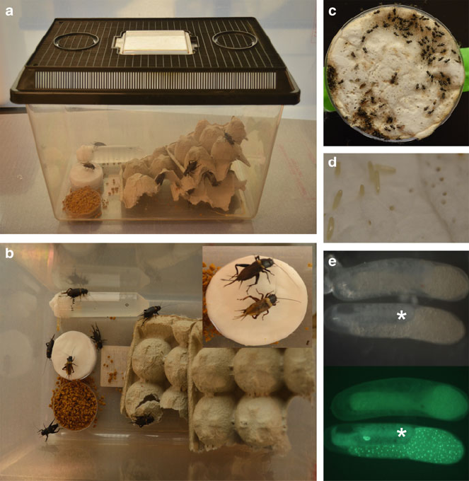
(Fig. 8.2 b ). Crickets will oviposit fertilized eggs into damp sand (e.g., Sandtastik
Sparkling White Play Sand, Product Code PLA0050), wet paper towels, Whatman
paper, or wet cotton placed in petri plates in cricket cages (Fig. 8.2b inset, d). These
eggs will develop successfully and hatch in 12–14 days (Fig. 8.2c) under the
following conditions: incubation at ~28–29
C with 70% humidity, dead or moldy
Fig. 8.2 G. bimaculatus husbandry. (a) (side view) and (b) (top view) showing a well-ventilated
plastic container used for housing a cricket colony. Note the use of egg cartons for providing shelter,
ground cat food, and a 50 ml Falcon filled with water and stopped with cotton as a food and water
source, respectively. A wet cotton plate (seen in a, b, and b inset) is placed in the adult cages for
females to oviposit fertilized eggs. Oviposited eggs (higher magnification shown in (d)) need to be
kept moist and clean until the eggs hatch. (c) A close-up of a cotton plate showing newly emerged
cricket hatchlings, which can then be transferred into new plastic cages with food, water, and shelter
until they reach adulthood. (e) Two G. bimaculatus eggs (6 days after egg laying) imaged under
bright field white light (top) and green florescent light (bottom). Both eggs are progeny obtained
from a cross between the histone2B-GFP (H2B-GFP) transgenic and wild-type G. bimaculatus line.
Embryos carrying the H2B-GFP transgene (bottom egg marked with white asterisk) can be
distinguished from non-transgenic embryos (top egg) based on the presence of bright florescent
nuclei, which is detectable from day 5 until day 10 after egg laying
190 A. Kulkarni and C. G. Extavour

embryo removal on a regular basis, and maintenance of a moist and clean substrate
(Mito and Noji 2008; Donoughe and Extavour 2016). Embryonic development for
this species has been divided into 16 stages based on morphological features of
eggs, developing embryos and their appendages (Niwa et al. 1997; Donoughe and
Extavour 2016). After hatching, nymphs undergo eight nymphal molts to finally
become adults over the next 5 weeks. The generation time (total time to adulthood
and sexual maturity) of G. bimaculatus is thus approximately 7 weeks at 29
C
(Fig. 8.3). Adults are thought to reach maximum fecundi ty 1 week after the final
molt (Mito and Noji 2008). Sexing and identification of virgin males and females are
straightforward: late-stage male and female nymphs can be separated based on the
presence or absence of an ovipositor and then isolated until they undergo the final
molt to sexual maturity (Fig. 8.1a). This also helps in setting up single mating
crosses, for example, to establish genetically modified lines. Similarly, precisely
timed egg collections are possible by placing egg collection petri dishes in the cages
and removing them at desired intervals (described in Donoughe and Extavour 2016).
Fig. 8.3 A schematic showing an overview of the G. bimaculatus life cycle. Selected embryonic
and nymphal developmental stages are shown, alongside the duration of each developmental stage
depicted in hours (orange arc and lines), days (green arc and lines), or weeks (blue arc and lines)
after egg laying. The colored arcs indicate the entire duration of time occupied by the indicated
developmental stage, whereas the lines show a cartoon schematic of the selected stages within this
time window. Each displayed embryonic stage during embryogenesis shows the position of the
embryo (in gray) relative to the yolk (yellow) within the egg and has a small description of features
that are characteristic of that developmental stage (depicted as egg stage “EgS”). Dotted lines with
arrowheads indicate the different movements that the embryo makes during the course of develop-
ment in this species. Upon hatching, nymphs undergo eight nymphal molts (indicated by solid black
arrows) to reach adulthood. Newly emerged adult animals are sexually mature at molting and begin
mating soon after. This figure is modified from Donoughe and Extavour (2016)
8 The Cricket Gryllus bimaculatus: Techniques for Quantitative .. . 191
A G. bimaculatus spontaneous mutant strain with white eyes (Fig. 8.1c–e) was
isolated by Isao Nakatani and colleagues at the University of Yamagata, Japan, in
1989 (referenced in Mito and Noji 2008). This is, to our knowledge, currently the
only available mutant, and its phenotype is caused by an autosomal recessive
mutation, referred to as gwhite (Niwa et al. 1997; Mito and Noji 2008). This mutant
is sometimes preferred for whole-mount gene expression analysis at late stages of
embryogenesis, owing to the fact that at this stage, the tissues are more transparent
compared to wild type (Niwa et al. 1997; Mito and Noji 2008).
8.4 Techniques for Quantitative and Functional Genetic
Analyses in G. bimaculatus
Here we discuss protocols and methodologies that have been established and are
currently available in the cricket G. bimaculatus, with the aim of making new users
aware of the plethora of techniques at their disposal. Detailed descriptions of these
published techniques and step-by-step protocols are thus avoided in this chapter
(we refer the reader to Horch et al. 2017a, b for detailed protocols). While these
protocols are now well established in crickets, these tools are not limited to them and
could in principle be modified or adapted for use in other hemimetabolous insects to
further species-specific research.
8.4.1 Precise Embryonic Staging System
To make meaningful observations of deviations from normal embryonic develop-
ment, one first needs a wild-type reference for any given species. Donoughe and
Extavour (2016) have reported a detailed embryonic staging system for
G. bimaculatus (Figs . 8.4 and 8.5). This system is based on externally observable
characters of the developing cricket embryo that are visible through the eggshell,
thereby circumventing the need for embryonic disse ctions to ascertain embryonic
developmental stage. This is especially informative for studying early embryos of
insects such as crickets, which are embedded within a large amount of opaque yolk,
making direct observations through the eggshell difficult if not impossible.
G. bimaculatus development, based on this staging, is presented as 24 “egg stages”
and encompasses the entire development of the animal from fertilization to hatching,
based solely on external observable egg characters (called stage identifiers). Each of
the 24 “egg stages” described here corresponds to one or more of 16 “ embryonic
stages.” For each stage, the authors provide a list of embryonic developmental
features defining that stage, including features of body segmentation, mesoderm,
and appendage formation. Determining the stage of embryogenesis through the
eggshell is a useful complement to earlier described staging schemes that require
dissection of the embryo (Mito and Noji 2008; Kainz 2009).
192 A. Kulkarni and C. G. Extavour

schematicbright field
6
7
9
10
11
12
1
2
8
egg
stage
(EgS)
embryonic
stage (ES)
lateral view of egg or
ventral view of embryo
1.0 – 1.4
1.6 – 4.0
4.0 – 5.2
5.2 – 6.5
7.5 – 8.5
7.0 – 7.5
6.0 – 7.0
1.5
7.2 – 8.0
8.7 – 9.5
3
4
5
8.0 – 8.7
8.5 – 9.0
Fig. 8.4 A detailed description of the egg stages (EgS 1–12) in G. bimaculatus. Micrographs in
second column from left help display the morphological features of the egg that can be used to
assign embryos to an egg stage (EgS) and also describe the corresponding morphological features
and embryonic stage (ES) of the embryo within the egg. In the schematic, the embryo is depicted in
gray and yolk in yellow. Micrographs in the right second column from the right are not to scale, and
are taken using lateral views of either a H2B-GFP transgenic live embryo (EgS 1–5) or ventral
views of dissected and fixed, Hoechst 33342-stained embryos (EgS 6-182). Micrographs are not to
scale. This figure is modified from Donoughe and Extavour (2016)
8 The Cricket Gryllus bimaculatus: Techniques for Quantitative .. . 193

schematicbright field
22
23
24
19
20
21
16
17
18
13
14
15
egg
stage
(ES)
embryonic
stage (ES)
lateral view of egg or
ventral view of embryo
9.0 – 9.9
11
11
11 – 12
13 – 14
12 – 13
11 – 12
10
12 – 14
16
14
15
Fig. 8.5 A detailed description of the egg stages (EgS 12–24) in G. bimaculatus. Staging system
for egg stages (EgS) 12–24 continued from Fig. 8.4. This staging system ends at hatching and does
not include postembryonic development of nymphs to adulthood. This figure is modified from
Donoughe and Extavour (2016)
194 A. Kulkarni and C. G. Extavour
8.4.2 Injection Methods for Eggs, Nymphs, and Adults
A basic requirement for many experimental procedures in modern developmental
biology, including live imaging, RNA interference, and gene editing, is the delivery
of synthetic or biological materials into the body of an animal, without disrupting its
health or sacrificing its life. Direct manual injection is one such method and, in the
case of crickets, has been well established and found effective in egg, nymphal,
and adult stages. Two methodological variations are commonly in use for
G. bimaculatus egg injections, diff ering essentially in the number and arrangement
of embryos for injection, and are described in great detail in Horch et al. (2017a) and
Barry et al. (2019). Both methods are thus described below in brief.
The first variant of the egg injection method, developed by the Noji lab (University
of Tokushima), involves the construction of a mold to house eggs for injections (Horch
et al. 2017a). Watson chambers (which resemble a rectangular mold) are glued onto
microscopic glass slides using double-sided tape. Embryos are then lined up end to end
along the length of the chamber, using a small stainless-steel spatula. The wall of the
chamber and the adhesive of the double-sided tape (on the slide) help secure and hold
the embryos in place during the injections. The second variant, optimized in the
Extavour lab, uses rectangular troughs made using plastic molds set in low-melting
agarose that hold eggs in place (Kainz et al. 2011; Barry et al. 2019). Using this setup,
over 35 embryos per slide can be prepared for injection simultaneously, making it
efficient in terms of preparation time and the number of embryos injected in one sitting.
Regardless of the egg injection method used, eggs are injected under a dissecting
or compound microscope, using a needle held by a micromanipulator (Fig. 8.6a, b).
The needle must be loaded with the desired injection material and connected to a
pressure source, which may be manual (e.g., a syringe) or electronically controlled
compressed gas (e.g., using a commercial micro-injector). The injected material may
be mixed with a dye that is visible under white light (e.g., phenol red or fast green) or
fluorescent light (e.g., fluorescein- or rhodamine-conjugated dextrans) depending on
the user’s preference. The choice of dye will determine the best microscopy and light
regime to be used for injections. Following injections, embryos are allowed to
develop normally in humid incubators at 28
C on wet paper towels or are sub-
merged in 1 phosphate buffer ed saline in closed petri dishes and monitored daily
until embryos hatch. Eggs of developmental stages that are turgid and under high
pressure, including very early stages in the first few hours following fertilization and
middle stages following elongation of the germ band, are more difficult to inject than
earlier stages.
For nymphal and adult injections, a manually held Hamilton syringe or any
automated micromanipulator/microinjector system specifically designed for delicate
microinjections and capable of injecting nanoliter volumes can be used effectively.
Nymphs and adults are prepared for injections by first cooling them on ice to
temporarily immobilize them, and then injected either between the abdominal
segments (A2 and A3) or in the soft tissue between the T3 coxa and the thorax.
For site- or tissue-specific injections (e.g., the leg or brain), the injection site should
8 The Cricket Gryllus bimaculatus: Techniques for Quantitative .. . 195

Fig. 8.6 Injection and OMMAwell setup. (a) The apparatus used for beveling glass needles for use
in injecting crickets, consisting of a light source, a dissecting microscope, a micromanipulator, and a
beveling stone (Narishige model EG-45 is shown here). (b) Higher magnification of the beveling
setup shown in (a). (c) Different types of agarose mold inserts used for mounting cricket embryos
for application in OMMAwell or, alternatively, for cricket egg injections. (d) OMMAwell
(Donoughe et al. 2018) schematic for top-loaded microwells, used for injecting and imaging cricket
embryos using a configuration for upright objectives. Different assembly components are shown:
the mold insert (white) consists of wells that will house the cricket embryos and is inverted and
attached to the base of the slide (gray). This is then placed into the upright platform (pink) and
196 A. Kulkarni and C. G. Extavour

be modified accordingly. However, locales of abundant fat tissue should be avoided
as injection sites in crickets, to help facilitate dispersal of liquid into the body upon
injection, prevent backflow of injected mat erial or hemolymph into the needle, and
prevent blockage of fine needle tips with insect tissue. Irrespective of the injection
site, care should be taken while injecting the needle into the nymphal or adult body,
so as to prevent injuring the internal organs, which cou ld potentially kill the animal
or disrupt recovery. Such injuries can be easily avoided by inserting the needle only
deep enough into the animal body to prevent oozing of material at injection.
Other recommendations for successful injections of adult or juvenile
G. bimaculatus include inserting the needle parallel to the body of the insect, rather
than at a perpendicular angle, injecting larger volumes (relative to the insect size) as
multiple pulses of smaller doses rather than all at once, injecting slowly to prevent
leakage, minimizing handling stress for the animal, making sure the needle is not
blocked prior to insertion, and maintaining basic cleanliness and sterility during the
procedure. Following injection, animals should be allowed to recover in isolated cages
with food and water at room temperature before proceeding with the desired study.
8.4.3 High-Throughput Live Imaging of Embryos Using
OMMAwell
Open Modular Mold for Agarose Microwells (OMMAwell) is a simple, reusable,
all-in-one device that allows users to easily mount and simultaneously image dozens
of live G. bimaculatus embryos consistently and economically for 2D and/or 3D
time-lapse analyses of early development (Donoughe et al. 2018). OM MAwell has
the added advantage of being adapta ble and customizable: it has been made to
accommodate the imaging needs of researchers with different experimental designs,
can be used on diverse species (OMMAwell has been successfully designed for and
tested on nine animal species, including many traditional model organisms), and can
be used for both inverted and upright objective microscopes (Fig. 8.6d). With this
device, embryos can be efficiently and quickly lined up in arrays of agarose
microwells, whose dimensions and spacing can also be customized as per individual
user needs (Fig. 8.6c) (see Donoughe et al. 2018). In addition, OMMAwell has
reservoirs to hold live imaging media and help maintain specimen-specific humidity,
osmolarity, and oxygen levels during time-lapse live imaging, thereby enhanci ng
embryonic survival and data quality. This device also allows positional tracking of
⁄
Fig. 8.6 (continued) secured at the desired height with the help of a pin (blue). The assembled
components are then lowered into a petri plate containing molten low-melt agarose and allowed to
set. Following the removal of the mold insert from the cooled and set agarose, specimens are added
into the wells in the petri plate either individually or in bulk and covered with low-melt agarose
(in microliter volumes of up to 100 μl) to hold them in place. Embryos can be oriented carefully
using forceps prior to this step. Once the agarose sets, live-imaging media is poured into the dish
8 The Cricket Gryllus bimaculatus: Techniques for Quantitative .. . 197
individual embryos and permits users to control samp le orientation for imaging. The
OMMAwell microwell array arrangement is also convenient to hold embryos in
place during injections. OMMAwell has been used to image the development of as
many as 102 G. bimaculatus live embryos simultaneously for 12 consecutive days
(Donoughe et al. 2018). Hatching rates of these embryos were not significantly
different from the hatching rates of controls, suggesting no lingering effects of
phototoxicity, developmental delays, or defects on these embryos from the use of
OMMAwell.
8.4.4 Gene Expression Analyses Using Embryonic or Whole
Mount In Situ Hybridization
and Immunohistochemistry
The ability to detect mRNAs [using in situ hybridization (ISH)] and proteins
[immunohistochemistry using label ed antibodies (IAb)] of interest is central to
whole mount gene expression analyses in any organism. Standard protocols to
study gene expression in other vertebrate and invertebrate embryos have been
applied successfully in G. bimaculatus (Niwa et al. 2000; Mito and Noji 2008).
These include whole mount in situ hybridizat ion using digoxigenin (DIG)-labeled
antisense RNA probes (as per Wilkinson 1992), protein detection (Patel 1994), and
double in situ hybridization using probes labeled with different haptens (e.g.,
Dietrich et al. 1997). Optimized ISH and IAb protocols have also been developed
in this species for specific tissues including the brain, nymphal legs, and wings.
Automated medium- or high-throughput gene expression assays on G. bimaculatus
tissues using specialized robots (e.g., Intavis InsituPro VSi) are also possible
(Extavour lab, unpublished).
8.4.5 RNA Interference
The Noji lab pioneered the establishment of RNA interference (RNAi) technology in
the cricket G. bimaculatus (Miyawaki et al. 2004), and many researchers have since
used this technique successfully to deplete mRNAs of multiple target genes in this
species. Four main types of RNAi techniques have been developed for use in crickets:
embryonic, nymphal, parental, and regenerative RNAi (Miyawaki et al. 2004;Mito
et al. 2005;Nakamuraetal.2008a; Mito and Noji 2008; Ronco et al. 2008).
To perform RNAi, double-stranded RNA (dsRNA), preferably 300–500 nucleo-
tides in length, complementary to a region in the G. bimaculatus gene of interest, is
injected into the eggs (embryonic RNAi) or into the body cavity of nymphs
(nymphal RNAi) or adults (paren tal RNAi). Successful concentrations of dsRNA
have been reported to range from 2 to 6 μg/μl (e.g., Kainz et al. 2011). It is
recommended that the dsRNAs designed should match a regio n close to or including
the 3
0
UTR of the target G. bimaculatus gene, which may minimize off-target effects.
Typical specificity controls may include testing at least one other dsRNA designed
198 A. Kulkarni and C. G. Extavour
against a nonoverlapping fragment of this same gene. Injecting dsRNA against
exogenous genes not encoded by the cricket genome (e.g., DsRed) and injecting
the buffer alone can also serve as meaningful controls and are thus stro ngly
recommended for every RNAi experiment. Together, these measures can help
researchers distingu ish between speci fic and nonspecific effects of RNAi, allowing
meaningful interpretation of their results.
RNAi is systemic in G. bimaculatus, such that RNAi-induced phenotypes may be
detected throughout the body of the embryo, nymph, or adult, regardless of the site
of injection. Moreover, the injection of dsRNA into sexually mature adult females
allows for observation of RNAi effects not only in the adult animal itself but also in
its progeny (i.e., eggs) that the animal will lay over the weeks following a
postinjection mating as long as the gene knockdown does not interfere with oogen-
esis, fertilization, or egg laying. Alternatively, nymphal RNAi can be conveniently
used to determine gene functions in postembryonic stages. Regenerative RNAi was
optimized in the Noji lab and has been performed as a specific application of
nymphal RNAi in the cricket (Nakamura et al. 2008a). For this procedu re, a leg of
a third instar nymph is amputated following dsRNA injection, and the effects of
RNAi are then assessed during the regeneration of the lost leg (which normally
occurs over subsequent molts). Based on these observations, the RNAi response in
crickets can be robust, stable, and even transmissible through subsequent molts
(Nakamura et al. 2008a; Hamada et al. 2015). However, it is recommended that
the robustness of RNAi response, its stability, and duration be determined on a case-
to-case basis; in our hands, there have been instances where the RNAi response for
some genes has lasted only a few days (see Kainz et al. 2011).
8.4.6 Calcium Imaging to Study Neurobiology
and Neuro ethology
The cricket has been an important model for neurobiology and neuroethological
studies, and many physiological techniques are easily applicable to the cricket
(Ogawa and Miller 2017). One such technique is that of calcium imaging, which
uses florescent dyes and optical methods to monitor the changes in intracellular
levels of calcium ions in live cells and tissues (Neubauer and MacLean 2010),
including cricket neurons. Information on selection of calcium indicators, dye
loading protocols, experimental desig ns, and calcium imaging techniques in the
cricket are well described (Ogawa and Miller 2017). In 2013, Matsumoto and
colleagues successfully expressed Yellow Chameleon (YC) 3.60, a genetically
encoded calcium indicator (GECI) in the cricket brain via electroporation
(Matsumoto et al. 2013), enabling prolonged deep imaging of the cricket brain for
the first time. Together with high-resolution microscopy and gene editing tech-
niques, calcium imaging is expected to facilitate major adv ances in our understand-
ing of cricket neurobiology (Ogawa and Miller 2017). Calcium imaging is not
limited to neurobiology, so its successful establishment in the study of cricket
8 The Cricket Gryllus bimaculatus: Techniques for Quantitative .. . 199
nervous tissue suggests that this technique can now also be used to study physiology
in other tissues and cell types in this animal.
8.4.7 High-Sensitivity Trackball Recording Systems
for Studying Phonotaxis and Auditory Neuronal
Plasticity
Acoustic communication is paramount in insects, both within and between species.
Making precise recordings of insect locomotory behavior in response to auditory
stimuli (phonotaxis), such as male calling songs, is often challenging under natural
settings. Various laboratory assays for measuring cricket phonotactic behavior have
been developed, making such studies possible. Examples of such assays include
analyzing the number of crickets that reach an acoustic stimuli or sound target within
adefined time period (Tschuch 1976; Stout et al. 1983), studying cricket behavior in
mazes (Popov and Shuvalov 1977; Rheinlaender and Blätgen 1982), or steering
responses of tethered flying female crickets (Pollack and Hoy 1979). The development
of two different trackball recording systems in crickets has been paramount in
enhancing our understanding of G. bimaculatus auditory steering behavior (Hedwig
2017) and provided detailed insights into insect locomotory behavior in general. All
trackball recording systems measure the movements of the trackball, based on which
insect velocity and direction of insect movement (walking) are inferred, without
allowing the insect to reach the auditory target. In closed-loop trackball systems, the
cricket is allowed to walk and turn freely on the trackball during recordings, with the
trackball compensating for cricket movement by having the ability to counter-rotate.
By contrast, in open-loop systems, the tethered cricket has the ability to walk but not
change its orientation in an acoustic field. Due to their sophisticated design, these
trackball recording systems can now easily be integrated into experimental setups
using other forms of recording, including neuro- or electrophysiological and high-
speed video recording experiments. Thus, combined with the GECI YC3.60 discussed
above, and alongside other sophisticated imaging and video recording techniques (see
below), trackball recording systems are expected to provide new insights not only into
cricket biology but also into the study of insect phonotaxis in general.
8.4.8 Automated and Customizable Video Tracking Systems,
Artificial Crickets, and Cricket Robots for Synthetic
Neuroethology and Social Behavior
Crickets have been used over the past several decades as systems to study behaviors
including mating, flight, aggression, wandering, obstacle avoidance, and importantly,
to study the neurophysiology underlying these processes. When investigating, quan-
tifying, and qualifying animal behavior, dependable and accurate measurement sys-
tems are needed to record animal responses to external stimuli, at both behavioral and
200 A. Kulkarni and C. G. Extavour
physiological levels. The advent of engineering approaches in crickets, especially
robotics, is expected to greatly facilitate such research and is the genesis of the field
of cricket synthetic neuroethology. Aonuma and colleagues have described a novel
approach developed for crickets, where provoked animal behavior in response to
computer-generated simulation and robots is captured to effectively bridge the gap
between insect behavior and physiology (Aonuma 2017). Different commercially
available automated video tracking systems designed to follow cricket movement
have also been previously described (Noldus et al. 2001). Recently, another custom-
izable tracking system based on a simple open-source solution called SwisTrack
(Lochmatter et al. 2008) has been introduced for use in crickets. Using this system,
multiple crickets can be video recorded and tracked simultaneously. Because the entire
process is semiautomatic, data collection and its interpretation are more efficient than
previous methods that were based exclusively on manual tracking. Using artificial
crickets (e.g., Funato et al. 2011; Kawabata et al. 2012;Mizunoetal.2012)orcricket
robots (Funato et al. 2008, 2011) alongside computer modeling is another way of
analyzing cricket behavior that has recently been reported. Further detailed informa-
tion on artificial crickets, cricket robots, biomimetic robots (Ritzmann et al. 2000), and
behavioral modeling in this species can be found in Aonuma (2017).
8.4.9 Standardized Protocols for Assessing Learning
and Me mory
G. bimaculatus has been reported to have a robust memory and thus has been
exploited for studying the ne ural mechanisms underlying olfactory, auditory, and
visual learning. Mizunami and colleagues have published detailed protocols for
classical conditioning, operant testing, associative learning, memory retention, and
subsequent data analyses in G. bimaculatus (Mizunami and Matsumoto 2017a). A
“classical conditioning and operant testing ” procedure has also been developed in
crickets by these researchers. The establishment of such protocols has resulted in the
elucidation of detailed cellular mechanisms and signaling cascades that are impor-
tant for memory formation in crickets. These studies have additionally revealed that
crickets display unexpected diversity in the mechanisms underlying these proces ses
in compa rison to other insects including Drosophila (Mizunami and Matsumoto
2017b). The use of such classical conditioning paradigms and their variants in
crickets may provide novel breakthroughs in our understanding of learning, cogni-
tion, and memory across animals.
8.4.10 Transgenic Lines
Stable transgenic lines are an invaluable tool for developmental genetics and con-
tribute to the successful establishment of a model animal system. Transgenesis using
P elements, which are the transposon of choice for Drosophila transgenesis (Rubin
8 The Cricket Gryllus bimaculatus: Techniques for Quantitative .. . 201
and Spradling 1982 ), have been found ineffective in crickets, such that other
transposable elements need to be used to achieve transformation in this species.
Zhang and colleagues (2002) showed that Minos transposons (Pavlopoulos et al.
2007) are active in G. bimaculatus embryos and highlighted the possibility of using
these as gene vectors for germ line transformation in this species. However, to our
knowledge, this transposon has not yet been used to establish stable transgenic
cricket lines. Shinmyo et al. (2004) succeeded in somatic transformation of
G. bimaculatus embryos, using the piggyBac transposon (Handler et al. 1998)to
achieve somatic insertion of a construct containing an enhanced green florescent
protein (eGFP) coding region driven by a G. bimaculatus actin 3/4 promoter.
Construction of plasmids and injection protocols for this line are as described in
Shinmyo et al. (2004) and Zhang et al. ( 2002). Subsequently, this technique has been
optimized to achieve germ line transmission of transgenes (Nakamura et al. 2010).
At the time of writing, a histone2B-GFP (H2B-GFP) transgenic line is stably
maintained in multiple laboratories (Nakamura et al. 2010 ). In this line, the promoter
of the G. bimaculatus actin orthologue (Gb-Actin) drives the expression of the
Gb-histone2B protein tagged with eGFP. This transgene is ubiquitously and consti-
tutively expressed and is maternally contributed to eggs. Based on viability ratios of
the embryos laid by this line, it is likely that the transgene is sublethal in homozy-
gosis (Extavour lab, unpublished observations). As there are no balancer chromo-
somes for G. bimaculatus, heterozygotes must be manually selected at every
generation to maintain the transgene (Extavour lab, unpublished observations).
Zygotic expression of this transgene begins at approximately the fourth day after
egg laying (AEL) at 28
C, and eggs expressing the transgene can then be easily
identified and selected between 5 and 10 days AEL, based on the presence of
brightly fluorescent nuclei under a fluorescent ster eomicroscope (Fig. 8.2e top and
bottom).
8.4.11 Genome Editing Using CRISPR/Cas9, TALE N,
and Zinc-Finger Nucleases
Sophisticated functional genetics techniques commonly used to modify genomes
in vivo at a specific site include clustered regularly interspaced palindromic repeats
(CRISPR)/CRISPR- associated nuclease 9 (Cas9), collectively known as the
CRISPR/Cas9 system (Cong et al. 2013), transcription activator-like (TAL) effector
nucleases (TALENs), and z inc-finger nucleases (ZFNs) (Porteus and Carroll 2005;
Moscou and Bogdanove 2009; Remy et al. 2010; Miller et al. 2011; Jinek et al.
2012). All of these approaches work by generating double-stranded breaks in target
DNA sequences, which in turn trigger the cell’s DNA damage response (Remy et al.
2010), and this cellular response can then generate mutations (insertions or dele-
tions) in the targeted gene. All of these techniques are now available and functional
in crickets. The Mito lab established and reported the use of ZFNs and TALENs in
202 A. Kulkarni and C. G. Extavour
crickets in 2012, by successfully creating homozygous genetic knockouts (Watanabe
et al. 2012). CRISPR/Cas9 has also now been effectively used for the generation of
both knock-ins (Horch et al. 2017b) and knockouts (Awata et al. 2015) of cricket
genes. Detailed protocols for knocking-in or knocking-out cricket genes using the
CRISPR/Cas9 method are available in Horch et al. (2017b).
8.4.12 Genomics and Transcriptomics
While no genome sequenc e is currently publicly available for G. bimaculatus,a
number of de novo transcriptomes have been published for this species, providing
gene expression datasets for a number of different specific tissue types and devel-
opmental stages. To date, these include transcriptomes of the ovaries, embryos, the
prothoracic ganglion, and regenerating legs (Zeng and Extavour 2012; Bando et al.
2013; Zeng et al. 2013; Fisher et al. 2018). Moreover, several transcriptomes
reflecting gene expression at different life stages (Berdan et al. 2016), in the male
accessory gland (Andres et al. 2013), fat body and flight muscles of different
ecological morphs (Vellichirammal et al. 2014), and under cold-acclimation condi-
tions (Des Marteaux et al. 2017; Toxopeus et al. 2019), are available for other
species of the genus.
8.5 Novel Insights into Biological Processes Using Forward
Genetics in a Hemimetabolous Insect
G. bimaculatus has been effectively used to study various disciplines of biological
sciences over the past decades. These include early embryonic development and
body patterning, tissue and organ system specification, regeneration, body size
regulation, memory and learning, reproductive biology, ecology, physiology, and
endocrinology (reviewed in Horch et al. 2017b). Here, we will therefore refrain from
reiterating the contributions that these resul ts have made to our understanding of
biology. Instead, we will briefly discuss one example of a novel insight that the
biological community has gained through the use of functional genetics in the
cricket.
8.6 The Evolution of the oskar Gene and Its Implications
for Germ-Line Research
Germ cells are the cells that give rise to eggs and sperm. They are therefore an
important cell type in sexually reproducing organisms and are sometimes referred to
as the ultimate totipotent stem cell (Cinalli et al. 2008), because they alone maintain
a genetic link between generations. In a developing embryo, the first cells to give rise
8 The Cricket Gryllus bimaculatus: Techniques for Quantitative .. . 203
to the germ cells by clonal mitoti c divisions are known as the primordial germ cells
(PGCs). Across metazoans, PGCs are speci fied using one of two mechanisms
(Extavour and Akam 2003). In some animals, including G. bimaculatus and Mus
musculus inductive cell–cell signaling among neighboring somatic cells instructs
certain cells to adopt PGC fate; this method of PGC specification is known as
“induction.” In other animals, including the fruit fly D. melanogaster, the nematode
worm Caenorhabditis elegans, the zebrafish Danio rerio, and the clawed frog
Xenopus laevis, PGCs are instead specified by “inheritance.” In this mechanism,
PGCs are specified very early in development through the cytoplasmic inheritance of
a maternally derive d special cytoplasm called “germ plasm,” which often contains
determinants that confer germ-line fate.
oskar is an insect-specific gene critical for the establishment of germ plasm in
D. melanogaster and is the only gene reported in the animal kingdom to be both
necessary and sufficient for germ cell formation (Lehmann and Nüsslein-Volhard
1986; Ephrussi et al. 1991; Kim-Ha et al. 1991; Ephrussi and Lehmann 1992; Smith
et al. 1992). Following its discovery in D. melanogaster, oskar orthologues were
reported in the genomes of other holometabolous insects known to specify their
germ cells using germ plasm (Goltsev et al. 2004; Juhn and James 2006; Juhn et al.
2008; Lynch et al. 2011). Interestingly, oskar appears to be absent from many insect
genomes that are known to lack germ plasm, including the bee A. mellifera, the
beetle T. castaneum, and the silk moth B. mori (summarized by Quan and Lynch
2016). Based on this observation and the fact that hemimetabolous insects reportedly
lack germ plasm (see Ewen-Campen et al. 2013 and references therein), it was
hypothesized that oskar was a novel gene that arose at the base of the Holometabola,
concurrent with the advent of insect germ plasm (Lynch et al.
2011). However,
Ewen-Campen and colleagues discovered an oskar orthologue in the cricket
G. bimaculatus genome and demonstrated that in this species, oskar is neither
expressed at high levels in PGCs nor required for PGC formation (Ewen-Campen
et al. 2012). Instead, cricket oskar is expressed in the neuroblasts (stem cells that
arise from the neural ectoderm and give rise to the nervous system in Pancrustacea)
of the brain and central nervous system (CNS) of the developing embryo, and
is required for proper embryonic CNS patterning (Ewen-Campen et al. 2012). This
observation, taken together with the reports that D. melanogaster oskar also plays a
neural role (Xu et al. 2013), suggests tw o novel hypotheses: (1) oskar arose at least
50 million years earlier in insect evolution than previously hypothesized, before the
divergence of Hemimetabola and Holometabola, and (2) oskar’s ancestral role in
insects may have been in the nervous system and not in the germ line. This implies
that oskar may have been co-opted for its essential role in holometab olous
germ plasm assembly rather than having originated concurrently with germ plasm
as had been previously sugges ted. This significantly changes our understanding of
the evolutionary origins and functional evolution not only of this gene but perhaps
also of insect germ plasm. Moreover, it constitutes an important example of how
novel genes may arise and become co-opted, across evolutionary time scales, to
perform different biological roles in animals.
204 A. Kulkarni and C. G. Extavour
8.7 Genomic Resources in Other Orthopterans
For an organism to become widely used as a research model for comparative or
evolutionary studies, an important contributing factor is whether resources and tools
are also available to study its close relatives. With this in mind, we will discuss
available resources in other orthopterans that may be of use in aiding comparative
work with G. bimaculatus. To our knowledge, at the time of writing, large-scale
genomic resources are available for only two other orthopterans, a locust and a
Hawaiian cricket species.
8.7.1 Resources in Field Crickets
Several transcriptomes are available for tissues and stages of many species of the
genus Gryllus, including (1) gene expression at different life stage s in G. rubens
(Berdan et al. 2016), (2) in the male accessory gland of G. firmus and
G. pennsylvanicus (Braswell et al. 2006; Andres et al. 2013), (3) fat body and flight
muscles of different ecological morphs of G. firmus (Vellichirammal et al. 2014),
(4) under cold-acclimation in G. veletis (Des Marteaux et al. 2017; Toxopeus et al.
2019), or (5) adult femur-derived transcriptomes from G. assimilis (Palacios-
Gimenez et al. 2018). Genomic resources are also available for an inbred line of
G. assimilis (Palacios-Gimenez et al. 2018). Outside of the genus Gry llus, large-
scale genomic resources are also available for the Hawaiian cricket Laupala
kohalensis in the form of an EST resource from a nerve cord cDNA library (Danley
et al. 2007) and a de novo draft genome (Blankers et al. 2018). In fact, the
L. kohalensis genome is, to our knowledge, the only published cricket genome to
date. Transcriptomic data are also a vailable for Allonemobius fasciatus embryos
(Reynolds and Hand 2009), male accessory glands of Gryllodes sigillatus (Pauchet
et al. 2015), and the testis, accessory glands, and adult body of Teleogryllus
oceanicus (Bailey et al. 2013).
8.7.2 Resources in Grasshoppers and Locusts
A de novo transcriptome spanning several stages is available for the grasshoppers
Chorthippus biguttulus and Oxya chinensis sinuosa (Kim et al. 2016) and for
nymphs, adult females and males of Xenocatantops brachycerus (Zhao et al.
2018). An organ-specific transcriptome is available for the gut of Oedaleus asiaticus
(Huang et al. 2017), an EST database exists for transcripts from the central nervous
system of Schistocerca gregaria (Badisco et al. 2011), and a de novo transcriptome
for Tetrix japonica (Qiu et al. 2017) is also available. In addition, other tools
including RNAi have been tested and reported to be successful in S. american a
8 The Cricket Gryllus bimaculatus: Techniques for Quantitative .. . 205
(Dong and Friedrich 2005). Locusta migratoria is a well-studied locust species that
has large-scale genomic resources available in the form of a de novo genome and
transcriptome (Wang et al. 2014) and an EST database from whole body and
dissected organs (Kang et al. 2004; Ma et al. 2006). In addition, RNAi has been
established and reported to be successful in adults, nymphs, and embryos for this
species (He et al. 2006).
8.8 Commercial Importance of Crickets as Edible Insects
and “Food of the Future”
In this chapter we have primarily focused on crickets as emerging evo-devo models.
In this final section, we would like to briefly highlight other reasons that crickets are
gaining popularity as study systems. Given their cosmopolitan distribution (all areas
of the world, except the arctic and subarctic regio ns) and over 2400 documented
species, crickets represent the most diverse lineage of “jumping or leaping” insects
(Horch et al. 2017b). While the chirping sounds made by males have historic ally
given them acoustic appeal as pets and in research, many species are now becoming
economically important as an alternative food source for human s (Huis et al. 2013;
Horch et al. 2017b), as feedstock for poultry (Ravindran and Blair 1993), or as fish
bait (Huis et al. 2013), all of which are multibillion dollar industries. With the human
population predicted to reach nine billion by the year 2050 (Huis et al. 2013), meat
production and consumption is soon expected to reach unsustainable levels (Boland
et al. 2013). Insects, especially crickets, have therefore been proposed and marketed
as a novel, alternative, environmentally efficient food source with high nutritional
value (Oonincx and de Boer 2012 ; Huis et al. 2013; Deroy et al. 2015).
Crickets are reportedly common street snacks in some parts of the world and have
been part of the traditional diet in Thailand, the Lao People’s Democratic Republic,
Vietnam, the Democratic Republic of Congo, and Nigeria for hundreds of years
(Kuhnlein et al. 2009; Huis et al. 2013). As an example, over 20,000 farmers are
reported to rear crickets in Thailand, resulting in an estimated production of over
7500 tons per year in this country alone (Hanboonsong et al. 2013). To increase their
appeal in the West, crickets are now being advertised and marketed to be eaten
whole, in granular or in paste form, and as ingredients in commercially available
flours and protein bars (e.g., Aspire Food Group USA, Inc.). While many species of
crickets are edible (e.g., G. bimaculatus, Gryllodes sigillatus, A. domesticus ,
A. testacea, T. occipitalis, T. mitratus, and Brachytrupes portentosus), to our
knowledge currently only G. bimaculatus, G. sigillatus, and A. domesticus are
farmed economically for human consumption (Huis et al. 2013).
The use of crickets has the potential to change the future of the food industry,
because of how effective they are in maximizing nutrition for minimal resources.
Cricket rearing is comparatively inexpensive, requires a fraction of input resources,
and has fewer negative environmental impacts than rearing traditional vertebrate
206 A. Kulkarni and C. G. Extavour
protein sources (Halloran et al. 2017). Crickets produce only 1% of greenhouse
gases compared to cattle and pigs, in addition to showing an approximate tenfold
reduction in ammonia emission (Oonincx et al. 2010), all relevant factors when
considering sustainable production in the age of clim ate change. They act as a
complete protein source and consi st of over 50% protein by volume (Wang et al.
2004). Other advantages of eating crickets include their high edible weight:
Nakagaki and DeFoliart (1991) have estimated that over 80% of a cricket is edible
and digestible compared to 55% for chicken and pigs and 40% for cattle. This
translates into making crickets twice as efficient as chicken, at least 4 times as
efficient as pigs, and 12 times more efficient as cattle in converting feed into meat
(Huis et al. 2013). As a specific example, the food conversion efficiency of the house
cricket Acheta domesticus has been reported to be five times higher than beef, and
when their fecundi ty is considered, this has been shown to increase as much as 15- to
20-fold (Horch et al. 2017b; Nakagaki and Defoliart 1991). Farming crickets is
projected to become a multimillion dollar industry, with the US market for edible
insects alone expected to exceed $50 million by as early as 2023 (Ahuja and Deb
2018).
The development of a novel food source like crickets must include assessing the
potential risks involved with consumption of such sources. Therefore, there is
increased interest in understanding the biology of these insects. While studies
addressing entomophagy-induced food allergies (especially ones arising from eating
crickets alone) are few, there is some preliminary evidence to confirm that
crustacean-allergic individuals (or people with seafood allergies) may also show
cross-reactivity to edible insects in general (Srinroch et al. 2015 ; Pener 2016; Ribeiro
et al. 2018). Another study has reported that some individuals can develop asthmatic
symptoms upon ingesting insects belonging to Orthoptera (Auerswald and Lopata
2005). Overall, however, eating and/or exposure to insects is not expected to pose
significant risks of allergenic reactions for most people, especially if the individual
has no prior history of arthropod or insect allergen sensiti vity (Huis et al. 2013). In
summary, the disadvantages associated with eating insects like crickets currently
seem few and the advantages many. Consuming reared insects is potentially more
environmentally friendly, nutritious, cheap, and affordable for people in all parts of
the world. Cricket rearing is one way to use land efficiently, reduce or lower
pesticide use and greenhouse gas emissions, may boost human and/or animal
immunity (Goodman 1989; Muzzarelli 2010; Taufek et al. 2016), and finally
improve the livelihood of women and children in rural areas by supporting local
economies (Huis et al. 2013).
8.9 Conclusion
The successful establishment of the many functional and genetic manipulation tools
in crickets has contributed to a new era of non-drosophilid insect research, not
limited to evo-devo research. We hope that scientists from various disciplines feel
8 The Cricket Gryllus bimaculatus: Techniques for Quantitative .. . 207
encouraged to use the cricket as a system to address intriguing questions in their
respective fields.
Acknowledgments We thank Extavour lab members Aracely Newton and Maitreyi Upadhyay for
helpful comments and Leo Blondel for technical support on the manuscript. This work was
supported by Harvard University.
References
Ahuja K, Deb S (2018) Edible insects market size by product (beetles, caterpillars, grasshoppers,
bees, wasps, ants, scale insects & true bugs), by application (flour, protein bars, snacks),
industry analysis report, Regional Outlook (U.S., Belgium, Netherlands, UK, France, China,
Thailand, Vietnam, Brazil, Mexico), application potential, price trends, competitive market
share & forecast, 2018– 2024
Alexander DE (2018) A century and a half of research on the evolution of insect flight. Arthropod
Struct Dev 47:322–327
Andres JA, Larson EL, Bogdanowicz SM, Harrison RG (2013) Patterns of transcriptome diver-
gence in the male accessory gland of two closely related species of field crickets. Genetics
193:501–513
Aonuma H (2017) Chapter 20: Synthetic approaches for observing and measuring cricket behav-
iors. In: Horch HW, Mito T, Popadic A, Ohuchi H, Noji S (eds) The cricket as a model
organism: development, regeneration and behaviour. Springer, New York
Auerswald L, Lopata AL (2005) Insects – diversity and allergy. Curr Allergy Clin Immunol
18:58–60
Awata H, Watanabe T, Hamanaka Y, Mito T, Noji S, Mizunami M (2015) Knockout crickets for the
study of learning and memory: dopamine receptor Dop1 mediates aversive but not appetitive
reinforcement in crickets. Sci Rep 5:15885
Badisco L, Huybrechts J, Simonet G, Verlinden H, Marchal E, Huybrechts R, Schoofs L, De
Loof A, Vanden Broeck J (2011) Transcriptome analysis of the desert locust central nervous
system: production and annotation of a Schistocerca gregaria EST database. PLoS One 6:
e17274
Bailey NW, Veltsos P, Tan YF, Millar AH, Ritchie MG, Simmons LW (2013) Tissue-specific
transcriptomics in the field cricket Teleogryllus oceanicus. G3 (Bethesda) 3:225–230
Bando T, Ishimaru Y, Kida T, Hamada Y, Matsuoka Y, Nakamura T, Ohuchi H, Noji S, Mito T
(2013) Analysis of RNA-Seq data reveals involvement of JAK/STAT signalling during leg
regeneration in the cricket Gryllus bimaculatus. Development 140:959–964
Barry S, Nakamura T, Matsuoka Y, Straub C, Horch HW, Extavour CG (2019) Injecting Gryllus
bimaculatus eggs. J Vis Exp (in press)
Berdan EL, Blankers T, Waurick I, Mazzoni CJ, Mayer F (2016) A genes eye view of ontogeny: de
novo assembly and profiling of the Gryllus rubens transcriptome. Mol Ecol Resour
16:1478–1490
Bigelow RS (1962) Factors affecting developmental rates and diapause in field crickets. Evolution
16:396–406
Bilinski SM, Jaglarz MK, Tworzydło W (2017) The pole (germ) plasm in insect oocytes. In: Kloc
M (ed) Results and problems in cell differentiation, oocytes, vol 63. Springer, New York, pp
103–126
Blankers T, Oh KP, Bombarely A, Shaw KL (2018) The genomic architecture of a rapid island
radiation: recombination rate variation, chromosome structure, and genome assembly of the
Hawaiian Cricket Laupala. Genetics 209:1329–1344
208 A. Kulkarni and C. G. Extavour
Boland MJ, Rae AN, Vereijken JM, Meuwissen MPM, Fischer ARH, Boekel MAJSv, Rutherfurd
SM, Gruppen H, Moughan PJ, Hendriks WH (2013) The future supply of animal-derived
protein for human consumption. Trends Food Sci Technol 29:62–73
Braswell WE, Andres JA, Maroja LS, Harrison RG, Howard DJ, Swanson WJ (2006) Identification
and comparative analysis of accessory gland proteins in Orthoptera. Genome 49:1069–1080
Bretman A, Tregenza T (2005) Measuring polyandry in wild populations: a case study using
promiscuous crickets. Mol Ecol 14:2169–2179
Bridges CB, Morgan TH (1923) The third-chromosome group of mutant characters of Drosophila
melanogaster. Carnegie Inst Wash Publ 327:1–251
Bruce HS, Patel NH (2018) Insect wings and body wall evolved from ancient leg segments.
bioRxiv, 244541
Büning J (1994) The insect ovary: ultrastructure, previtellogenic growth and evolution. Chapman &
Hall, London
Campos-Ortega JA, Hartenstein V (1985) The embryonic development of Drosophila
melanogaster. Springer, Heidelberg
Cinalli RM, Rangan P, Lehmann R (2008) Germ cells are forever. Cell 132:559–562
Cong L, Ran FA, Cox D, Lin S, Barretto R, Habib N, Hsu PD, Wu X, Jiang W, Marraffini LA,
Zhang F (2013) Multiplex genome engineering using CRISPR/Cas systems. Science
339:819–823
Danley PD, Mullen SP, Liu F, Nene V, Quackenbush J, Shaw KL (2007) A cricket gene index: a
genomic resource for studying neurobiology, speciation, and molecular evolution. BMC Geno-
mics 8:109
Davis GK, Patel NH (2002) Short, long, and beyond: molecular and embryological approaches to
insect segmentation. Annu Rev Entomol 47:669–699
Demerec M (1950) Biology of Drosophila, Facsimile edn. Cold Spring Harbor Laboratory, Cold
Spring Harbor, NY
Denell R (2008) Establishment of Tribolium as a genetic model system and its early contributions to
evo-devo. Genetics 180:1779–1786
Deroy O, Reade B, Spence C (2015) The insectivore’s dilemma, and how to take the West out of
it. Food Qual Prefer 44:44–55
Des Marteaux LE, McKinnon AH, Udaka H, Toxopeus J, Sinclair BJ (2017) Effects of cold-
acclimation on gene expression in fall field cricket (Gryllus pennsylvanicus) ionoregulatory
tissues. BMC Genomics 18:357
Dietrich S, Schubert FR, Lumsden A (1997) Control of dorsoventral pattern in the chick paraxial
mesoderm. Development 124:3895–3908
Dong Y, Friedrich M (2005) Nymphal RNAi: systemic RNAi mediated gene knockdown in
juvenile grasshopper. BMC Biotechnol 5:25
Donoughe S, Extavour CG (2016) Embryonic development of the cricket Gryllus bimaculatus. Dev
Biol 411:140–156
Donoughe S, Kim C, Extavour CG (2018) High-throughput live-imaging of embryos in microwell
arrays using a modular specimen mounting system. Biol Open 7:bio031260
Duboule D, Dolle P (1989) The structural and functional organization of the murine HOX gene
family resembles that of Drosophila homeotic genes. EMBO J 8:1497–1505
Engel JE, Hoy RR (1999) Experience-dependent modification of ultrasound auditory processing in
a cricket escape response. J Exp Biol 202:2797–2806
Ephrussi A, Lehmann R (1992) Induction of germ cell formation by oskar. Nature 358:387–392
Ephrussi A, Dickinson LK, Lehmann R (1991) Oskar organizes the germ plasm and directs
localization of the posterior determinant nanos
. Cell 66:37–50
Ewen-Campen B, Srouji JR, Schwager EE, Extavour CG (2012) Oskar predates the evolution of
germ plasm in insects. Curr Biol 22:2278–2283
Ewen-Campen B, Donoughe S, Clarke DN, Extavour CG (2013) Germ cell specification requires
zygotic mechanisms rather than germ plasm in a basally branching insect. Curr Biol 23:835–842
8 The Cricket Gryllus bimaculatus: Techniques for Quantitative .. . 209
Extavour CG, Akam ME (2003) Mechanisms of germ cell specification across the metazoans:
epigenesis and preformation. Development 130:5869–5884
Ferreira M, Ferguson JW (2010) Do Mediterranean crickets Gryllus bimaculatus De Geer (Orthop-
tera: Gryllidae) come from the Mediterranean? Large-scale phylogeography and regional gene
flow. Bull Entomol Res 100:49–58
Fisher HP, Pascual MG, Jimenez SI, Michaelson DA, Joncas CT, Quenzer ED, Christie AE, Horch
HW (2018) De novo assembly of a transcriptome for the cricket Gryllus bimaculatus protho-
racic ganglion: an invertebrate model for investigating adult central nervous system compensa-
tory plasticity. PLoS One 13:e0199070
Funato T, Kurabayashi D, Nara M, Aonuma H (2008) Switching mechanism of sensor-motor
coordination through an oscillator network model. IEEE Trans Syst Man Cybern B Cybern
38:764–770
Funato T, Nara M, Kurabayashi D, Ashikaga M, Aonuma H (2011) A model for group-size-
dependent behaviour decisions in insects using an oscillator network. J Exp Biol
214:2426–2434
Geer CD (1773) Mémoires pour servir a l’histoire des insectes. Pierre Hesselberg, Stockholm
Giribet G, Edgecombe GD (2013) The Arthropoda: a phylogenetic framework. In: Minelli A,
Boxshall G, Fusco G (eds) Arthropod biology and evolution – molecules, development,
morphology. Springer, New York, pp 17–40
Goldsmith MR, Shimada T, Abe H (2005) The genetics and genomics of the silkworm, Bombyx
mori. Annu Rev Entomol 50:71–100
Goltsev Y, Hsiong W, Lanzaro G, Levine M (2004) Different combinations of gap repressors for
common stripes in Anopheles and Drosophila embryos. Dev Biol 275:435–446
Goodman WG (1989) Chitin: a magic bullet? The Food Insects Newsletter 2
Gould JL, Grould CG (1995) The honey bee. Scientific American Library, New York
Graham A, Papalopulu N, Krumlauf R (1989) The murine and Drosophila homeobox gene
complexes have common features of organization and expression. Cell 57:367–378
Grimaldi D, Engel MS (2005) Evolution of the insects. Cambridge University Press, Cambridge
Halloran A, Hanboonsong Y, Roos N, Bruun S (2017) Life cycle assessment of cricket farming in
north-eastern Thailand. J Clean Prod 156:83–94
Hamada Y, Bando T, Nakamura T, Ishimaru Y, Mito T, Noji S, Tomioka K, Ohuchi H (2015) Leg
regeneration is epigenetically regulated by histone H3K27 methylation in the cricket Gryllus
bimaculatus. Development 142:2916–2927
Hanboonsong Y, Jamjanya T, Durst PB (2013) Six-legged livestock: edible insect farming,
collection and marketing in Thailand. Food and Agriculture Organization of the United Nations
Regional Office for Asia and the Pacific, Bangkok
Handler AM, McCombs SD, Fraser MJ, Saul SH (1998) The lepidopteran transposon vector,
piggyBac, mediates germ-line transformation in the Mediterranean fruit fly. Proc Natl Acad
Sci USA 95:7520–7525
Harvey TH, Velez MI, Butterfield NJ (2012) Exceptionally preserved crustaceans from western
Canada reveal a cryptic Cambrian radiation. Proc Natl Acad Sci USA 109:1589–1594
He ZB, Cao YQ, Yin YP, Wang ZK, Chen B, Peng GX, Xia YX (2006) Role of hunchback in
segment patterning of Locusta migratoria manilensis revealed by parental RNAi. Dev Growth
Differ 48:439–445
Hedwig B (2017) Chapter 19: Trackball systems for analysing cricket phonotaxis. In: Horch HW,
Mito T, Popadic A, Ohuchi H, Noji S (eds) The cricket as a model organism: development,
regeneration and behaviour. Springer, New York
Hedwig B, Poulet JF (2004) Complex auditory behaviour emerges from simple reactive steering.
Nature 430:781–785
Heffer A, Xiang J, Pick L (2013) Variation and constraint in Hox gene evolution. Proc Natl Acad
Sci USA 110:2211–2216
210 A. Kulkarni and C. G. Extavour
Horch HW, Liu J, Mito T, Popadic A, Watanabe T (2017a) Chapter 21: Protocols in the cricket. In:
Horch HW, Mito T, Popadic A, Ohuchi H, Noji S (eds) The cricket as a model organism:
development, regeneration and behaviour. Springer, New York, pp 327–370
Horch HW, Mito T, Popadic A, Ohuchi H, Noji S (2017b) The cricket as a model organism:
development, regeneration and behaviour. Springer, New York, p 376
Huang X, Ma J, Qin X, Tu X, Cao G, Wang G, Nong X, Zhang Z (2017) Biology, physiology and
gene expression of grasshopper Oedaleus asiaticus exposed to diet stress from plant secondary
compounds. Sci Rep 7:8655
Huber F, Moore TE, Loher W (1989) Cricket behavior and neurobiology. Cornell University Press,
Ithaca, NY
Huis Av, Itterbeeck JV, Klunder H, Mertens E, Halloran A, Muir G, Vantomme P (2013) Edible
insects: future prospects for food and feed security. Food and Agriculture Organization of the
United Nations, Rome, p 201
Jinek M, Chylinski K, Fonfara I, Hauer M, Doudna JA, Charpentier E (2012) A programmable
dual-RNA-guided DNA endonuclease in adaptive bacterial immunity. Science 337:816–821
Juhn J, James AA (2006) oskar gene expression in the vector mosquitoes, Anopheles gambiae and
Aedes aegypti. Insect Mol Biol 15:363–372
Juhn J, Marinotti O, Calvo E, James AA (2008) Gene structure and expression of nanos (nos) and
oskar (osk) orthologues of the vector mosquito, Culex quinquefasciatus. Insect Mol Biol
17:545–552
Kainz F (2009) Cell communication during patterning: Notch and FGF signalling in Gryllus
bimaculatus and their role in segmentation. Department of Zoology, University of Cambridge,
Cambridge, p 161
Kainz F, Ewen-Campen B, Akam M, Extavour CG (2011) Delta/Notch signalling is not required for
segment generation in the basally branching insect Gryllus bimaculatus. Development
138:5015–5026
Kang L, Chen X, Zhou Y, Liu B, Zheng W, Li R, Wang J, Yu J (2004) The analysis of large-scale
gene expression correlated to the phase changes of the migratory locust. Proc Natl Acad Sci
USA 101:17611–17615
Kawabata K, Fujii T, Aonuma H, Suzuki T, Ashikaga M, Ota J, Asama H (2012) A
neuromodulation model of behavior selection in the fighting behavior of male crickets. Robot
Auton Syst 60:707–713
Kijimoto T, Pespeni M, Beckers O, Moczek AP (2013) Beetle horns and horned beetles: emerging
models in developmental evolution and ecology. Wiley Interdiscip Rev Dev Biol 2:405–418
Kim IW, Markkandan K, Lee JH, Subramaniyam S, Yoo S, Park J, Hwang JS (2016) Transcriptome
profiling and in silico analysis of the antimicrobial peptides of the grasshopper oxya chinensis
sinuosa. J Microbiol Biotechnol 26:1863 –1870
Kim-Ha J, Smith JL, Macdonald PM (1991) oskar mRNA is localized to the posterior pole of the
Drosophila oocyte. Cell 66:23–35
Kleespies RG, Tidona CA, Darai G (1999) Characterization of a new iridovirus isolated from
crickets and investigations on the host range. J Invertebr Pathol 73:84–90
Kochi Y, Miyashita A, Tsuchiya K, Mitsuyama M, Sekimizu K, Kaito C (2016) A human
pathogenic bacterial infection model using the two-spotted cricket, Gryllus bimaculatus.
FEMS Microbiol Lett 363:fnw163
Kochi Y, Matsumoto Y, Sekimizu K, Kaito C (2017) Two-spotted cricket as an animal infection
model of human pathogenic fungi. Drug Discov Ther 11:259–266
Krause G (1939) Die Eitypen der Insekten. Biol Zentralbl 50:495–536
Kuhnlein HV, Erasmus B, Spigelski D (2009) Indigenous Peoples
’ food systems: the many
dimensions of culture, diversity and environment for nutrition and health. Food and Agriculture
Organization of the United Nations Center for Indigenous Peoples’ Nutrition and Environment,
Rome
8 The Cricket Gryllus bimaculatus: Techniques for Quantitative .. . 211
Lehmann R, Nüsslein-Volhard C (1986) Abdominal segmentation, pole cell formation, and embry-
onic polarity require the localized activity of oskar, a maternal gene in Drosophila. Cell
47:144–152
Lewis EB (1978) A gene complex controlling segmentation in Drosophila. Nature 276:565–570
Linz DM, Tomoyasu Y (2018) Dual evolutionary origin of insect wings supported by an investi-
gation of the abdominal wing serial homologs in Tribolium. Proc Natl Acad Sci U S A 115:
E658–E667
Liu PZ, Kaufman TC (2005) Short and long germ segmentation: unanswered questions in the
evolution of a developmental mode. Evol Dev 7:629–646
Lochmatter T, Roduit P, Cianci CM, Correll N (2008) SwisTrack – a flexible open source tracking
software for multi-agent systems. In: IEEE/RSJ international conference on intelligent robots
and systems, Acropolis Convention Center, Nice
Lohs-Schardin M, Cremer C, Nusslein-Volhard C (1979) A fate map for the larval epidermis of
Drosophila melanogaster: localized cuticle defects following irradiation of the blastoderm with
an ultraviolet laser microbeam. Dev Biol 73:239–255
Lynch JA, Özüak O, Khila A, Abouheif E, Desplan C, Roth S (2011) The phylogenetic origin of
oskar coincided with the origin of maternally provisioned germ plasm and pole cells at the base
of the Holometabola. PLoS Genet 7:e1002029
Ma Z, Yu J, Kang L (2006) LocustDB: a relational database for the transcriptome and biology of the
migratory locust (Locusta migratoria). BMC Genomics 7:11
Matsumoto CS, Shidara H, Matsuda K, Nakamura T, Mito T, Matsumoto Y, Oka K, Ogawa H
(2013) Targeted gene delivery in the cricket brain, using in vivo electroporation. J Insect Physiol
59:1235–1241
Mayhew PJ (2007) Why are there so many insect species? Perspectives from fossils and phylog-
enies. Biol Rev Camb Philos Soc 82:425–454
Meng X, Zhu F, Chen K (2017) Silkworm: a promising model organism in life science. J Insect Sci
17:97, 1–6
Miller JC, Tan S, Qiao G, Barlow KA, Wang J, Xia DF, Meng X, Paschon DE, Leung E, Hinkley
SJ, Dulay GP, Hua KL, Ankoudinova I, Cost GJ, Urnov FD, Zhang HS, Holme s MC, Zhang L,
Gregory PD, Rebar EJ (2011) A TALE nuclease architecture for efficient genome editing. Nat
Biotechnol 29:143–148
Mitchell AA (2013) EDNA. The Fossil Insect Database. https://fossilinsectdatabase.co.uk/
Mito T, Noji S (2008) The two-spotted cricket Gryllus bimaculatus: an emerging model for
developmental and regeneration studies. Cold Spring Harbor Protoc. https://doi.org/10.1101/
pdb.emo110
Mito T, Sarashina I, Zhang H, Iwahashi A, Okamoto H, Miyawaki K, Shinmyo Y, Ohuchi H, Noji S
(2005) Non-canonical functions of hunchback in segment patterning of the intermediate germ
cricket Gryllus bimaculatus. Development 132:2069–2079
Mito T, Nakamura T, Bando T, Ohuchi H, Noji S (2011) The advent of RNA interference in
entomology. Entomol Sci 14:1–8
Miyawaki K, Mito T, Sarashina I, Zhang H, Shinmyo Y, Ohuchi H, Noji S (2004) Involvement of
Wingless/Armadillo signaling in the posterior sequential segmentation in the cricket, Gryllus
bimaculatus (Orthoptera), as revealed by RNAi analysis. Mech Dev 121:119–130
Mizunami M, Matsumoto Y (2017a) Chapter 9: Leaning and memory. In: Horch HW, Mito T,
Popadic A, Ohuchi H, Noji S (eds) The cricket as a model organism: development, regeneration
and behaviour. Springer, New York, pp 129–140
Mizunami M, Matsumoto Y (2017b) Roles of octopamine and dopamine neurons for mediating
appetitive and aversive signals in Pavlovian conditioning in crickets. Front Physiol 8:1027
Mizuno T, Sakura M, Ashikaga M, Aonuma H, Chiba R, Ota J (2012) Model of a sensory–
behavioral relation mechanism for aggressive behavior in crickets. Robot Auton Syst
60:700–706
Moscou MJ, Bogdanove AJ (2009) A simple cipher governs DNA recognition by TAL effectors.
Science 326:1501
212 A. Kulkarni and C. G. Extavour
Muzzarelli RAA (2010) Chitins and chitosans as immunoadjuvants and non-allergenic drug
carriers. Mar Drugs 8:292–312
Nakagaki BJ, Defoliart GR (1991) Comparison of diets for mass-rearing Acheta domesticus
(Orthoptera: Gryllidae) as a novelty food, and comparison of food conversion efficiency with
values reported for livestock. J Econ Entomol 84:891–896
Nakamura T, Mito T, Bando T, Ohuchi H, Noji S (2008a) Dissecting insect leg regeneration
through RNA interference. Cell Mol Life Sci 65:64–72
Nakamura T, Mito T, Miyawaki K, Ohuchi H, Noji S (2008b) EGFR signaling is required for
re-establishing the proximodistal axis during distal leg regeneration in the cricket Gryllus
bimaculatus nymph. Dev Biol 319:46–55
Nakamura T, Yoshizaki M, Ogawa S, Okamoto H, Shinmyo Y, Bando T, Ohuchi H, Noji S, Mito T
(2010) Imaging of transgenic cricket embryos reveals cell movements consistent with a syncy-
tial patterning mechanism. Curr Biol 20:1641–1647
Neubauer FB, MacLean JN (2010) Calcium imaging in neuroscience. In: Encycopedia of Life
Sciences (ELS). John Wiley & Sons, Ltd, Chichester
Nicholson DB, Ross AJ, Mayhew PJ (2014) Fossil evidence for key innovations in the evolution of
insect diversity. Proc Biol Sci 281:20141823
Nickle DA, Walker TJ (1974) A morphological key to field crickets of southeastern United States
(Orthoptera: Gryllidae: Gryllus). Fla Entomol 57:8–12
Niwa N, Saitoh M, Ohuchi H, Yoshioka H, Noji S (1997) Correlation between distal-less expres-
sion patterns and structures of appendages in development of the two-spotted cricket, Gryllus
bimaculatus. Zool Sci 14:115–125
Niwa N, Inoue Y, Nozawa A, Saito M, Misumi Y, Ohuchi H, Yoshioka H, Noji S (2000)
Correlation of diversity of leg morphology in Gryllus bimaculatus (cricket) with divergence
in dpp expression pattern during leg development. Development 127:4373–4381
Noldus LP, Spink AJ, Tegelenbosch RA (2001) EthoVision: a versatile video tracking system for
automation of behavioral experiments. Behav Res Methods Instrum Comput 33:398–414
Nüsslein-Volhard C, Wieschaus EF (1980) Mutations affecting segment number and polarity in
Drosophila. Nature 287:795–801
Ogawa H, Miller JP (2017) Chapter 18: Optical recording methods: how to measure neural
activities with calcium imaging. In: Horch HW, Mito T, Popadic A, Ohuchi H, Noji S (eds)
The cricket as a model organism: development, regeneration and behaviour. Springer,
New York
Oldroyd BP, Thompson GJ (2006) Behavioural genetics of the honey bee Apis mellifera. Adv
Insect Physiol 33:1–49
Oonincx DG, de Boer IJ (2012) Environmental impact of the production of mealworms as a protein
source for humans – a life cycle assessment. PLoS One 7:e51145
Oonincx DG, van Itterbeeck J, Heetkamp MJ, van den Brand H, van Loon JJ, van Huis A (2010) An
exploration on greenhouse gas and ammonia production by insect species suitable for animal or
human consumption. PLoS One 5:e14445
Otte D, Cade W (1984) African crickets (Gryllidae). 5. East and South African species of
Modicogryllus and several related genera (Gryllinae, Modicogryllini). Proc Acad Natl Sci
Phila 136:67–97
Palacios-Gimenez OM, Bardella VB, Lemos B, Cabral-de-Mello DC (2018) Satellite DNAs are
conserved and differentially transcribed among Gryllus cricket species. DNA Res 25:137–147
Panganiban G, Irvine SM, Lowe C, Roehl H, Corley LS, Sherbon B, Grenier JK, Fallon JF,
Kimble J, Walker M, Wray GA, Swalla BJ, Martindale MQ, Carroll SB (1997) The origin
and evolution of animal appendages. PNAS 94:5162–5166
Patel NH (1994) Imaging neuronal subsets and other cell types in whole-mount Drosophila
embryos and larvae using antibody probes. Methods Cell Biol 44:445–487
Pauchet Y, Wielsch N, Wilkinson PA, Sakaluk SK, Svatos A, ffrench-Constant RH, Hunt J, Heckel
DG (2015) What
’s in the gift? Towards a molecular dissection of nuptial feeding in a cricket.
PLoS One 10:e0140191
8 The Cricket Gryllus bimaculatus: Techniques for Quantitative .. . 213
Pavlopoulos A, Oehler S, Kapetanaki MG, Savakis C (2007) The DNA transposon Minos as a tool
for transgenesis and functional genomic analysis in vertebrates and invertebrates. Genome Biol
8(Suppl 1):S2
Paydar S, Doan CA, Jacobs GA (1999) Neural mapping of direction and frequency in the cricket
cercal sensory system. J Neurosci 19:1771–1781
Pener MP (2016) Allergy to crickets: a review. J Orthop Res 25:91–95
Pollack GS, Hoy RR (1979) Temporal pattern as a cue for species-specific calling song recognition
in crickets. Science 204:429 –432
Popov AV, Shuvalov VF (1977) Phonotactic behavior of crickets. J Comp Physiol 119:111–126
Porteus MH, Carroll D (2005) Gene targeting using zinc finger nucleases. Nat Biotechnol
23:967–973
Qiu Z, Liu F, Lu H, Huang Y (2017) Characterization and analysis of a de novo transcriptome from
the pygmy grasshopper Tetrix japonica. Mol Ecol Resour 17:381–392
Quan H, Lynch JA (2016) The evolution of insect germline specification strategies. Curr Opin
Insect Sci 13:99–105
Ravindran V, Blair R (1993) Feed resources for poultry production in Asia and the Pacific. III.
Animal protein sources. Worlds Poult Sci J 49:219–235
Remy S, Tesson L, Menoret S, Usal C, Scharenberg AM, Anegon I (2010) Zinc-finger nucleases: a
powerful tool for genetic engineering of animals. Transgenic Res 19:363–371
Reynolds JA, Hand SC (2009) Embryonic diapause highlighted by differential expression of
mRNAs for ecdysteroidogenesis, transcription and lipid sparing in the cricket Allonemobius
socius. J Exp Biol 212:2075–2084
Rheinlaender J, Blätgen G (1982) The precision of auditory lateralization in the cricket, Gryllus
bimaculatus. Physiol Entomol 7:209–218
Ribeiro JC, Cunha LM, Sousa-Pinto B, Fonseca J (2018) Allergic risks of consuming edible insects:
a systematic review. Mol Nutr Food Res 62:1700030
Ritzmann RE, Quinn RD, Watson JT, Zill SN (2000) Insect walking and biorobotics: a relationship
with mutual benefits. Bioscience 50:23– 33
Ronco M, Uda T, Mito T, Minelli A, Noji S, Klingler M (2008) Antenna and all gnathal appendages
are similarly transformed by homothorax knock-down in the cricket Gryllus bimaculatus. Dev
Biol 313:80–92
Rubin GM, Spradling AC (1982) Genetic transformation of Drosophila with transposable element
vectors. Science 218:348–353
Sander K (1997) Pattern formation in insect embryogenesis: the evolution of concepts and mech-
anisms. Int J Insect Morphol Embryol 25:349–367
Sen GL, Blau HM (2006) A brief history of RNAi: the silence of the genes. FASEB J 20:1293–1299
Shinmyo Y, Mito T, Matsushita T, Sarashina I, Miyawaki K, Ohuchi H, Noji S (2004) piggyBac-
mediated somatic transformation of the two-spotted cricket, Gryllus bimaculatus. Dev Growth
Differ 46:343–349
Simmons LW (1986) Female choice in the field cricket
Gryllus bimaculatus (De Geer). Anim
Behav 34:1463–1470
Simmons LW (1987) Female choice contributes to offspring fitness in the field cricket, Gryllus
bimaculatus (De Geer). Behav Ecol Sociobiol 21:313–321
Smith JL, Wilson JE, Macdonald PM (1992) Overexpression of oskar directs ectopic activation of
nanos and presumptive pole cell formation in Drosophila embryos. Cell 70:849–859
Sokoloff A (1966) The genetics of Tribolium and related species. Academic Press, New York
Sokoloff A (1972) The biology of Tribolium with special emphasis on genetic aspects, vol 1.
Clarendon, Oxford
Sokoloff A (1974) The biology of Tribolium with special emphasis on genetic aspects, vol 2.
Clarendon, Oxford
Sokoloff A (1977) The biology of Tribolium with special emphasis on genetic aspects, vol 3.
Clarendon, Oxford
214 A. Kulkarni and C. G. Extavour
Srinroch C, Srisomsap C, Chokchaichamnankit D, Punyarit P, Phiriyangkul P (2015) Identification
of novel allergen in edible insect, Gryllus bimaculatus and its cross-reactivity with
Macrobrachium spp. allergens. Food Chem 184:160–166
Stout JF, DeHaan CG, McGhee RW (1983) Attractiveness of the male Acheta domesticus calling
song to females. J Comp Physiol 153:509–521
Szelei J, Woodring J, Goettel MS, Duke G, Jousset FX, Liu KY, Zadori Z, Li Y, Styer E, Boucias
DG, Kleespies RG, Bergoi n M, Tijssen P (2011) Susceptibility of North-American and
European crickets to Acheta domesticus densovirus (AdDNV) and associated epizootics. J
Invertebr Pathol 106:394–399
Taufek NM, Aspani F, Muin H, Raji AA, Razak SA, Alias Z (2016) The effect of dietary cricket
meal (Gryllus bimaculatus) on growth performance, antioxidant enzyme activities, and
haematological response of African catfish (Clarias gariepinus). Fish Physiol Biochem
42:1143–1155
Toth AL, Rehan SM (2017) Molecular evolution of insect sociality: an eco-evo-devo perspective.
Annu Rev Entomol 62:419– 442
Toxopeus J, Des Marteaux LE, Sinclair BJ (2019) How crickets become freeze tolerant: the
transcriptomic underpinnings of acclimation in Gryllus veletis. Comp Biochem Physiol Part D
Genomics Proteomics 29:55–66
Tregenza T, Wedell N (1998) Benefits of multiple mates in the cricket Gryllus bimaculatus.
Evolution 52:1726–1730
Tregenza T, Wedell N (2002) Polyandrous females avoid costs of inbreeding. Nature 415:71–73
Truman JW, Riddiford LM (1999) The origins of insect metamorphosis. Nature 401:447–452
Tschuch G (1976) Der Einfluss synthetischer Gesänge auf die Weibchen von Gryllus bimaculatus
De Geer. Zool Jahrb Abt allgemeine Zool Physiol Tiere 80:383–388
Vaccari NE, Edgecombe GD, Escudero C (2004) Cambrian origins and affinities of an enigmatic
fossil group of arthropods. Nature 430:554–557
Vellichirammal NN, Zera AJ, Schilder RJ, Wehrkamp C, Riethoven J-JM, Brisson JA (2014) De
novo transcriptome assembly from fat body and flight muscles transcripts to identify morph-
specific gene expression profiles in Gryllus firmus. PLoS One 9:e82129
Wang Y, Jehle JA (2009) Nudiviruses and other large, double-stranded circular DNA viruses of
invertebrates: new insights on an old topic. J Invertebr Pathol 101:187–193
Wang D, Bai Y-Y, Li J-H, Zhang C-X (2004) Nutritional value of the field cricket (Gryllus
testaceus Walker). Entomol Sin 11:275–283
Wang X, Fang X, Yang P, Jiang X, Jiang F, Zhao D, Li B, Cui F, Wei J, Ma C, Wang Y, He J,
Luo Y, Wang Z, Guo X, Guo W, Wang X, Zhang Y, Yang M, Hao S, Chen B, Ma Z, Yu D,
Xiong Z, Zhu Y, Fan D, Han L, Wang B, Chen Y, Wang J, Yang L, Zhao W, Feng Y, Chen G,
Lian J, Li Q, Huang Z, Yao X, Lv N, Zhang G, Li Y, Wang J, Wang J, Zhu B, Kang L (2014)
The locust genome provides insight into swarm formation and long-distance
flight. Nat
Commun 5:2957
Watanabe T, Ochiai H, Sakuma T, Horch HW, Hamaguchi N, Nakamura T, Bando T, Ohuchi H,
Yamamoto T, Noji S, Mito T (2012) Non-transgenic genome modifications in a hemimetabo-
lous insect using zinc- finger and TAL effector nucleases. Nat Commun 3:1017
Wenzel B, Hedwig B (1999) Neurochemical control of cricket stridulation revealed by pharmaco-
logical microinjections into the brain. J Exp Biol 202:2203–2216
Werren JH, Loehlin DW (2009) The parasitoid wasp Nasonia: an emerging model system with
haploid male genetics. Cold Spring Harb Protoc. https://doi.org/10.1101/pdb.emo134
Wheeler QD (1990) Insect diversity and cladistic constraints. Ann Entomol Soc Am 83:1031–1047
Wilkinson DG (1992) In situ hybridization: a practical approach. Oxford University Press, Oxford
Xia Q, Zhou Z, Lu C, Cheng D, Dai F, Li B, Zhao P, Zha X, Cheng T, Chai C, Pan G, Xu J, Liu C,
Lin Y, Qian J, Hou Y, Wu Z, Li G, Pan M, Li C, Shen Y, Lan X, Yuan L, Li T, Xu H, Yang G,
Wan Y, Zhu Y, Yu M, Shen W, Wu D, Xiang Z, Yu J, Wang J, Li R, Shi J, Li H, Li G, Su J,
Wang X, Li G, Zhang Z, Wu Q, Li J, Zhang Q, Wei N, Xu J, Sun H, Dong L, Liu D, Zhao S,
Zhao X, Meng Q, Lan F, Huang X, Li Y, Fang L, Li C, Li D, Sun Y, Zhang Z, Yang Z, Huang Y,
8 The Cricket Gryllus bimaculatus: Techniques for Quantitative .. . 215
Xi Y, Qi Q, He D, Huang H, Zhang X, Wang Z, Li W, Cao Y, Yu Y, Yu H, Li J, Ye J, Chen H,
Zhou Y, Liu B, Wang J, Ye J, Ji H, Li S, Ni P, Zhang J, Zhang Y, Zheng H, Mao B, Wang W,
Ye C, Li S, Wang J, Wong GK, Yang H (2004) A draft sequence for the genome of the
domesticated silkworm (Bombyx mori). Science 306:1937–1940
Xu X, Brechbiel JL, Gavis ER (2013) Dynein-dependent transport of nanos RNA in Drosophila
sensory neurons requires rumpelstiltskin and the germ plasm organizer oskar. J Neurosci
33:14791–14800
Yoshimura A (2005) Karyotypes of two American field crickets: Gryllus rubens and Gryllus
sp. (Orthoptera: Gryllidae). Entomol Sci 8:219 –222
Zeng V, Extavour CG (2012) ASGARD: an open-access database of annotated transcriptomes for
emerging model arthropod species. Database. https://doi.org/10.1093/database/bas048
Zeng V, Ewen-Campen B, Horch HW, Roth S, Mito T, Extavour C (2013) Developmental gene
discovery in a hemimetabolous insect: de novo assembly and annotation of a transcriptome for
the cricket Gryllus bimaculatus. PLoS One 8:e61479
Zhang H, Shinmyo Y, Hirose A, Mito T, Inoue Y, Ohuchi H, Loukeris TG, Eggleston P, Noji S
(2002) Extrachromosomal transposition of the transposable element Minos in embryos of the
cricket Gryllus bimaculatus. Dev Growth Differ 44:409–417
Zhang XG, Maas A, Haug JT, Siveter DJ, Waloszek D (2010) A eucrustacean metanauplius from
the lower Cambrian. Curr Biol 20:1075–1079
Zhao L, Zhang X, Qiu Z, Huang Y (2018) De novo assembly and characterization of the
Xenocatantops brachycerus transcriptome. Int J Mol Sci 19:520
216 A. Kulkarni and C. G. Extavour
